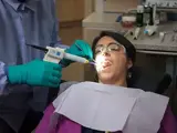
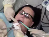
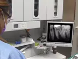
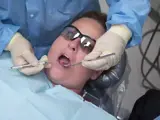
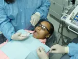
This module is part of a 2-volume set. Part I of this module explains the dental assistant’s role in dental radiography procedures; including bitewing and periapical full mouth series radiographs, utilizing the paralleling technique. The paralleling technique is the most commonly used technique for exposing periapical and bitewing radiographs because it creates the most accurate representation of a tooth image. It refers to the receptor being positioned parallel to the full length (long axis) of the tooth being radiographed. The central x-ray beam is then aligned perpendicular to the tooth being radiographed. Film holding devices, such as the XCP (extension-cone paralleling) device are explained in this module, along with the 5 basic rules for performing the paralleling technique.
Dental Radiography 1 - Parallel Technique
Dental Assisting
Materials Included

Text

Video

Anatomy

Simulation

Quiz
This module is part of a 2-volume set. Part I of this module explains the dental assistant’s role in dental radiography procedures; including bitewing and periapical full mouth series radiographs, utilizing the paralleling technique.
